(一)目前的多重检测技术主要是检测显色染料、金属同位素、荧光基团等发出的信号来达到检测标志物的目的。各个mIHC/IF平台的工作原理图(Diagram showing mechanism of each of the mIHC/IF platform)如图1所示:
图1A. DISCOVERY ULTRA system : af ter pr ima ry ant ibo dy incubati on, a second ary an tibody label led with HRP is intr oduc ed. The H RP is reac ted wi th an a ppropri at e su bs tr at e bo un d to a ch romo geni c d ye, le adi ng t o th e pr ec ip it at io n of i n s o l u b l e , c o l o u r e d p r e c i p i t a t e s a t t h e s i t e w h e r e t h e a n t i g e n s a r e f o u n d .
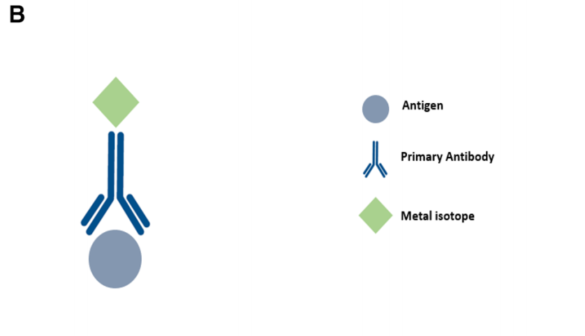
图1B. Metal-based IHC techniques such as IMC and MIBI: a pr ima ry ant ibo dy bou nd to the ta rget a ntigen is ta gged with a m etal iso tope of know n mo lecul ar ma ss. A nalysi s is ca rried o ut u si ng m as s sp ec tr om et ry in MIB I a nd las er a bl at io n co up le d to m as s c y t o m e t r y i n I M C .
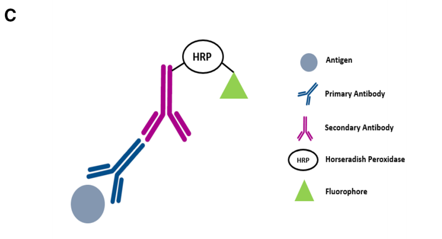
图1C. Vectra : aft er pri mar y a nti bod y incuba tion, a seco ndary antibo dy l abel led with HRP is intr oduc ed. A fluo ropho re-con jugated t yr am id e mo le cu le s er ve s as the sub str ate fo r HR P, r es ul ti ng i n an a nt ig e n - a s s o c i a t e d f l u o r e s c e n c e s i g n a l .
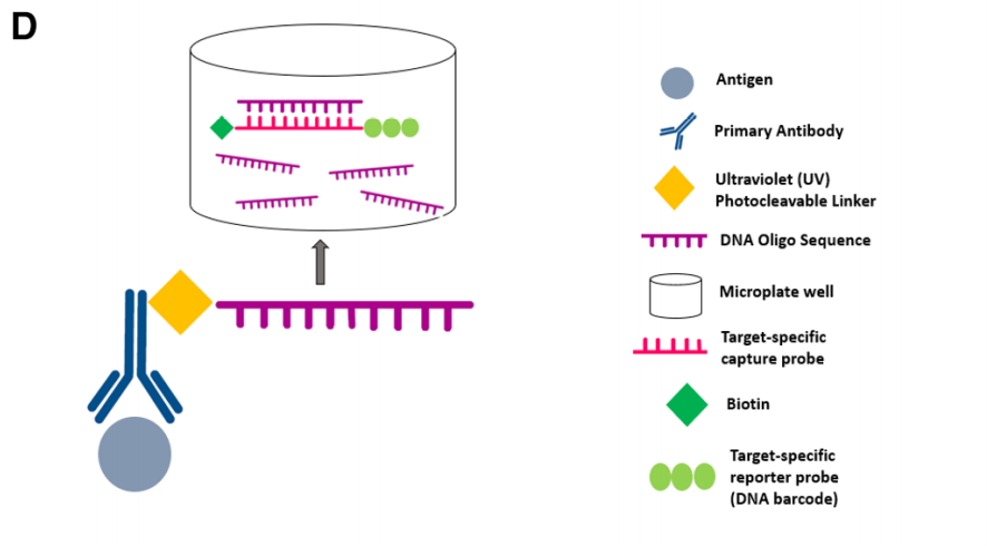
图1D. Nanostring’s DSP : the ta rge t a nti gen will bind the p rimary antib ody wh ich is coupl ed t o a phot o cl eava ble olig onucl eotid e tag . UV l ight is u se d to c le av e th e ol ig on ucle otid e t ags an d is c ol le ct ed u si ng a m ic ro c a p i l l a r y t u b e a n d s t o r e d i n a m i c r o p l a t e w e l l . T h e o l i g o n u c l e o t i d e t a g s w i l l b i n d t o t h e r e p o r t e r p r o b e v i a t h e t a r g e t - s p e c i f i c c a p t u r e p r o b e . R e p o r t e r p r o b e s a r e i m a g e d a n d c o u n t e d b y t h e n C o u n t e r a n a l y s i s s y s t e m .
(二)现有的多重检测技术可同时检测5-40+标志物,不同成像平台的比较(Overview and comparison of the different imaging modalities)如表1所示:
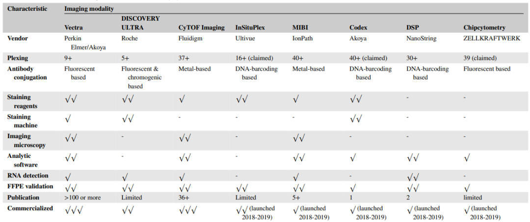
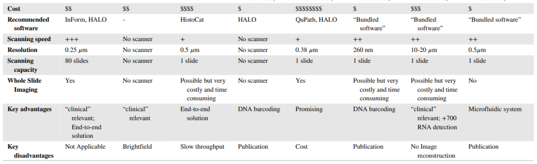
表1. Overview and comparison of the different imaging modalities
(三)Vectra, Chipcytometry, or DISCOVERY ULTRA平台mIHC/IF图像示例(Representative mIHC/IF images captured through the Vectra, Chipcytometry, or DISCOVERY ULTRA imaging system):
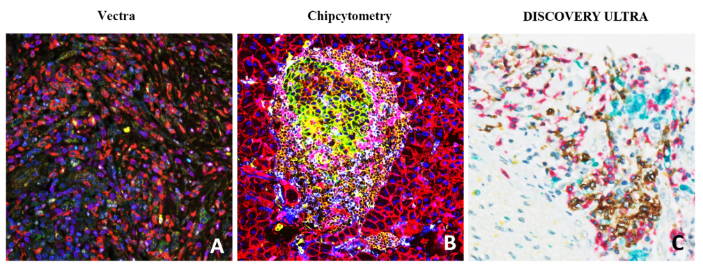
图2. Representative mIHC/IF images captured through the Vectra, Chipcytometry, or DISCOVERY ULTRA imaging system.
-
mIHC/IF of pancreatic adenocarcinoma FFPE sections labelled with DAPI (blue), CD73 (green), CD8 (yellow), CD68 (red), FoxP3 (cyan), CD3(magenta) and CK (orange) were scanned using the Vectra imaging system.
-
Mouse pancreas FFPE sections labelled with CD45 (brown), CD274(green), CD3e (purple), CD4 (cyan), CD8a (pink), CD11b (yellow), CD31 (darkbrown), CD326/EpCAM (red), B220 (orange), F4/80 (blue), NK1.1 (purple), Pan-CK(maroon), Hoechst 33342 (dark blue) were scanned using the Chip cytometry imaging system.
-
Cholangiocarcinoma FFPE sections labelled with CD20 (blue), CD8(red), CD68 (turquoise), CD3 (yellow) were scanned using the DISCOVERY ULTRA imaging system.
(四)IMC技术最大的优势在于可同时检测40+标志物,图3为人体组织切片IMC图像示例(Representative IMC images of human tissue sections):
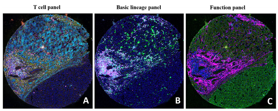
图3. Each image depicts the tumor microenvironment with the following immune cell lineages:
-
T cell panel
(A; C D 4 5 R O d e p i c t e d i
ngr
e e n
, C
K i n cya n ,
col l ag e
n i n y
ell o w ,
C D8 i n r e d
, C D4 i n m a ge n ta , and K i 67 i n w
h i t e)
-
Basic lineage panel (B; CD68 in green, CD20 in cyan, PD-L1 in yellow, VISTA in red, CD3 in magenta, and CD45 in white)
-
Function panel (C; OX40 in green, CD38 in cyan, Ki67 in yellow, ecadherin in red, collagen in magenta, and granzymeB in white).
(五)mIHC/IF图像分析仍然面临很多挑战,现有的解决方案各有优劣势,其中HistoCAT、QuPath属于开源的分析软件,常用的mIHC/IF 图像分析平台(Some of the commonly adopted analytic platforms for mIHC/IF)优劣势比较如下:
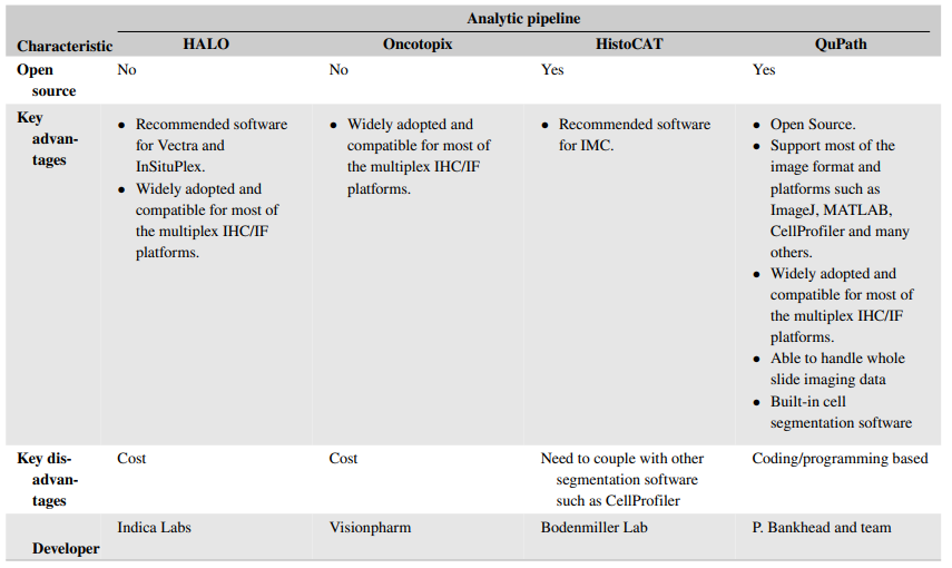
表2. Some of the commonly adopted analytic platforms for mIHC/IF
(六)Ultivue’s InSituPlex可使用普通的荧光显微镜成像,不需要配置昂贵的显微镜,因此可在大部分实验室开展检测。图4为人体组织样本Ultivue’s InSituPlex图像示例(Representative Ultivue’s InSituPlex images of human tissue samples labelled with CD8 (green), CD68 (yellow), PD-L1 (red) and CK/Sox10 (cyan)):
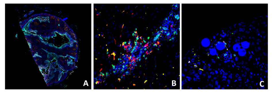
图4. Representative Ultivue’s InSituPlex images of human tissue samples labelled with CD8 (green), CD68 (yellow), PD-L1 (red) and CK/Sox10(cyan). Whole slide imaging of tonsil section (A), high magnification view ofHCC (B), and radioembolization-treated HCC (C, Y-90 visible as microspheres).
(七)目前已研究过多种mIHC/IF Panel,通过同时分析多个标志物来预测患者接受PD-1/PD-L1抑制剂治疗的应答反应。Meta分析中使用mIHC/IF技术的文献清单(List of papers using mIHC/IF in the meta-analysis)如下:
表3. List of papers using mIHC/IF in the meta-analysis
综上所述,mIHC/IF在肿瘤免疫治疗领域具有广阔的应用前景。与传统的IHC只能检测一个标志物不同,mIHC/IF能够在单个组织切片中检测多个标志物,同时提供有关细胞组成和空间排列的全面信息,使我们能够更深入地了解癌症的发病机制和对免疫治疗的反应。同时mIHC/IF技术处理的组织样本可以长期保存,供进一步研究使用。但是,这类技术的检测成本及实用性方面,仍然是一个值得关注的问题。
迈杰转化医学作为国内精准诊断整体解决方案的领导者,致力于解决精准医疗药物研发及患者用药痛点,围绕生物标志物研究、伴随诊断开发,建立了完善的核酸组学、蛋白组学、细胞组学技术平台。我们拥有国内领先的IHC检测平台,配备有Leica Bond Max、Ventana BenchMark、Dako Autostainer Link48三大进口自动化平台,以及用于mIHC检测的Leica Bond RX、PerkinElmerVectra3 System平台,在mIHC检测方法学开发及验证方面积累了丰富的经验,如三标四色的Panel(CD4, CD8, PD-L1 ),及五标六色的Panel(CD4, CD8, CD163, PD-L1, Pan-Keratin)等,如有mIHC检测方法开发及服务需求,请联系迈杰转化医学商务部(邮箱:
MARKETING@MEDxTMC.com
)。
Overview of multiplex immunohistochemistry/immunofluorescence techniques in the era of cancer immunotherapy - Tan - 2020 - Cancer Communications - Wiley Online Library(https://onlinelibrary.wiley.com/doi/10.1002/cac2.12023)
原创:Heisenberg
编辑:Grace


 关注微信公众号
 English
有限公司")